The Hidden Network Inside You That Could Change How You Think About Fertility and Red Light Therapy
If you have been trying to conceive for a while, I imagine you have heard some version of this sentence: "Everything looks fine." Your bloodwork is normal. Your scans are normal. Your partner's results are normal. And yet here you are, month after month, wondering why "normal" is not translating into a pregnancy.
I hear this from women in my practice all the time, and it is one of the most frustrating places to sit. You are doing everything right on paper, but your body has not gotten the memo.
So when something like pelvic red light therapy comes along a device you use at home, with suggestions that it might support your ovaries it makes sense that you would want to understand it before you invest your time, money, or hope in it. And more than that, you deserve to understand what is actually happening inside your body when you use it.
That is what this post is for. I am going to walk you through some genuinely fascinating anatomy that most women — and even many practitioners — have never been told about.
In This Article
What Pelvic Red Light Therapy Actually Is
Pelvic red light therapy, sometimes called intravaginal photobiomodulation, uses a small device inserted into the vagina that emits red, near-infrared (NIR) (and sometimes blue light). It is a form of low-level light therapy similar in principle to the red light panels used for skin health or muscle recovery, but designed specifically for the tissue inside the vaginal canal.
The idea behind photobiomodulation is not new. It has been studied for decades in wound healing, muscle recovery, and skin conditions. What is newer is the application to reproductive health, and specifically the question researchers and clinicians are now exploring: can this kind of light, delivered from inside the body, help create a better environment for conception?
The answer requires understanding a part of your anatomy that you were almost certainly never taught about in school and that most women only discover exists when something goes wrong.
Can Light Really Reach My Ovaries?
This is the question I get asked most often, and I want to answer it honestly, because you deserve a straight answer rather than a marketing one.
No. Light cannot physically travel from inside your vagina all the way to your ovaries. Your ovaries sit somewhere between 6 and 10 centimeters away from the vaginal canal, and red and near-infrared light does not travel in straight lines through the body the way a flashlight beam travels through air. As LED light moves through tissue, it scatters and gets absorbed, and the amount of light remaining drops off dramatically with every centimeter of depth. Laser reaches deeper tissues before scattering. By the time you account for the vaginal wall, connective tissue, and the distance to the ovary, there is no meaningful number of photons reaching the ovary directly. If anyone tells you this device is "zapping your eggs," that is not how physics works, and you should be wary of that claim.
So does that mean the whole idea falls apart? Not quite. It means the real mechanism is more interesting, and more indirect than "shining a light on the target."
The Real Mechanism: Your Body's Own Messaging System
Here is the part that actually matters, and I want to explain it in simple terms.
Every cell in your body has mitochondria, tiny structures inside the cell that produce energy. Think of them as the cell's rechargeable battery packs. Inside those mitochondria is an enzyme called cytochrome c oxidase, which is particularly good at absorbing red and near-infrared light. When light of the right wavelength hits tissue that is close enough to the surface to actually receive it, in this case, the vaginal mucosa and the tissue around the cervix it activates this enzyme. Those mitochondria start working more efficiently, producing more energy.
But here is the key insight: those locally-stimulated cells do not just sit there feeling better. They release signalling molecules into the bloodstream, things like nitric oxide (a natural blood vessel relaxer), ATP (cellular energy currency), and tiny packets of cellular information called exosomes. Think of these as text messages sent out into your circulation, rather than a light beam physically travelling to a distant organ.
This is called a systemic signalling effect, or in research contexts, a bystander effect, a local event that triggers a response somewhere else in the body, carried entirely through the bloodstream.
In July 2025, researchers led by Glen Jeffery published a study in Scientific Reports that demonstrated exactly this principle in a different part of the body. Participants were exposed to 850 nanometre near-infrared light on their torso, with their eyes fully shielded. Their colour vision improved within 24 hours, even though no light ever touched their eyes. Only 0.004% of the original light made it through the body, yet that tiny fraction was enough to trigger mitochondrial signalling that produced a measurable biological change in an organ the light never reached. The photochemical event was local. The biological response was systemic.
That is precisely the principle at work with intravaginal light therapy and the ovarian environment.
Pelvic red light therapy to optimize reproductive signalling
Let Me Introduce You to the Most Important Structure You Have Never Heard Of
Now we get to the fascinating part, the anatomy that makes intravaginal light delivery uniquely positioned compared to anything applied to the skin.
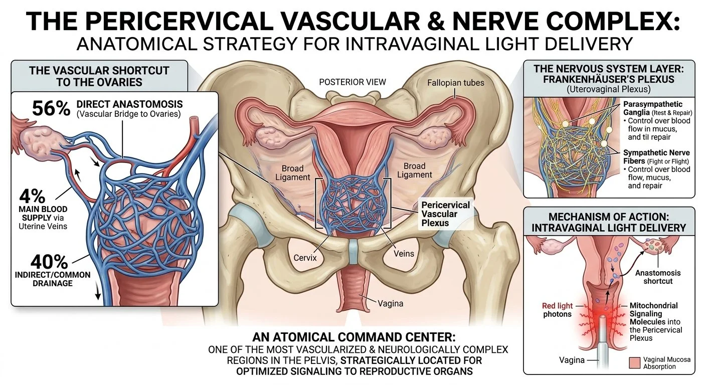
Sitting around your cervix is a structure called the pericervical vascular plexus. Let me explain what both of those words mean, because once you understand it, you will not forget it.
A plexus is simply a network, like a tangle of roads that all converge at the same junction. Not one road, not two roads, but many roads meeting at a single busy interchange. In your body, a vascular plexus is a dense tangle of blood vessels, veins and sometimes arteries clustered together in one area. The pericervical vascular plexus is that tangle of veins surrounding your cervix, on either side of it, within the broad ligament (the sheet of tissue that holds your uterus in place).
Picture it this way: imagine your cervix is a busy underground train station. The pericervical plexus is every platform, every tunnel, every connecting corridor all converging at that station at once. Blood from your vaginal walls, your cervix, your lower uterus, all of it funnels through this junction before heading back toward the heart.
Now here is what makes this even more fascinating.
The Bridge Between Your Uterus and Your Ovaries
In 56% of women, the pericervical venous plexus is physically connected to the ovarian blood supply through what is called an anastomosis. An anastomosis is simply a direct connection between two blood vessels, a biological bridge, like a canal connecting two rivers. It means blood (and whatever is dissolved in it including those signalling molecules from light-activated mitochondria) does not have to travel through the general circulation and hope it finds its way to the ovary. In more than half of all women, there is a direct shortcut.
In a smaller group (around 4% of women), the uterine venous system is actually the main blood supply to the ovaries, not the ovarian veins at all.
For the remainder of women, the ovarian veins are the primary route, and the connection is less direct. But even then, both systems drain into the same pelvic venous network, so signalling molecules entering the pericervical plexus are still entering a circulation that is closely intertwined with the ovarian environment.
This anatomy is why intravaginal light delivery is not just convenient — it is anatomically strategic. The vaginal mucosa feeds directly into the pericervical plexus. The pericervical plexus connects, in the majority of women, directly to the ovarian blood supply. It is like being able to drop something into the exact river that flows to where you need it, rather than releasing it into the ocean. Think of a message in a bottle…dropping that message into a river instead of the Atlantic ocean makes it more likely the message will reach it’s destination.
The Nervous System Layer - Two Systems in One Place
The pericervical area does not just carry blood vessels. Running right alongside those vessels is one of the most complex nerve networks in the female pelvis, the uterovaginal plexus, also called Frankenhäuser's plexus after the 19th-century German gynaecologist who first described it (not 100% sure if that’s a great name for it…).
This nerve network carries both the sympathetic nervous system (your "fight or flight" signals, which among other things control blood vessel tone) and the parasympathetic nervous system (your "rest and repair" signals, which regulate secretory activity, cervical mucus, and reflex responses). It is the control center for the uterus, cervix, and vagina — the final relay station that tells these organs what to do.
This means the pericervical area is simultaneously one of the most vascularised and one of the most neurologically complex regions in the entire female pelvis. It is not a passive pipe. It is an active command center sitting at the intersection of your circulatory and autonomic nervous systems.
Understanding Uterine Motility — And Why It Matters More Than Most Women Know
Before we talk about what can go wrong in this area, I need to introduce you to a concept that most women have never heard of: uterine motility.
When people think of the uterus contracting, they think of labor. But your uterus is moving all the time, long before pregnancy, long before labor. The muscle layer of the uterus (called the myometrium) produces gentle, rhythmic, wave-like contractions throughout your cycle. These are not painful in the way labor contractions can be. You likely never feel them. But they are doing critical work.
Before ovulation, these contractions are more frequent and directed upward, toward the fallopian tubes. Their job is to help transport sperm from the cervix up toward the egg waiting to be fertilised. Think of it like the uterus generating a gentle current to help sperm swim in the right direction.
After ovulation, once fertilization may have occurred, those contractions slow down significantly. The uterus becomes quieter, almost still. This is not a malfunction. It is deliberate. A calm, quiet uterus is what allows a fertilized egg (now a blastocyst) to travel to the right spot, make contact with the uterine lining, and begin to implant. Too many contractions at this stage, or contractions moving in the wrong direction, are associated with lower implantation success.
The autonomic nerve network in the pericervical area - Frankenhäuser's plexus is a key part of what controls this motility. Sympathetic nerve fibres regulate the timing and direction of those contractions. When those nerves are functioning well, the uterus moves correctly at the right time. When they are not, the rhythm is off. There is SO much more to implantation.
https://blog.tracydonegan.org/blog/red-light-therapy-and-implantation-success
https://blog.tracydonegan.org/blog/red-light-therapy-and-implantation-success
Gynae Procedures That Can Quietly Disrupt This System
This is the section I most want women to read, because almost no one talks about it.
The cervical nerve bundles that form part of Frankenhäuser's plexus are arranged in 12–16 individual nerve bundles within the cervix itself. They are physically present in the tissue that is accessed during many common gynaecological procedures. And research now shows that several of these procedures, often described as minor, routine, or low-risk can damage or disrupt these nerves in ways that are not immediately obvious but may have long-term consequences for fertility and uterine function.
Here are the procedures most associated with potential pericervical nerve disruption:
Dilation and curettage (D&C) - performed after miscarriage, for heavy bleeding, or to investigate the uterine lining. The process of dilating the cervix stretches the cervical tissue and can stress or injure the nerve bundles running through it. Vigorous curettage (scraping of the uterine lining) can additionally disrupt the nerve plexus at the endometrial-myometrial layer.
Application of a vulsellum clamp - a multi-toothed instrument applied to the front of the cervix to hold it in place during procedures. Research has shown that this can injure both layers of cervical nerve bundles simultaneously. The effects may not appear immediately, studies suggest a latent period of approximately three years before consequences become apparent, at which point women may experience painful hysteroscopy or difficult IUD insertion, without ever connecting these to the earlier procedure.
Cervical traction - excessive pulling on the cervix during any procedure can stretch or avulse (tear away) the nerve bundles running alongside the uterine vessels. Research has associated this with impaired fertility and reduced uterine motility.
Uterine evacuation procedures - including surgical management of miscarriage or termination. The normal nerve plexus at the junction of the uterine lining and muscle is vulnerable during these procedures. Repeated surgical evacuation is associated with adenomyosis (a condition where the uterine lining grows into the muscle wall), and the nerve disruption is thought to be part of that pathway.
LEEP (Loop Electrosurgical Excision Procedure) and cone biopsy - both used to remove abnormal cervical cells. While most research finds that the direct effect on fertility is relatively limited, the electrical current and tissue removal involved do affect the tissue architecture of the cervix, including the nerves passing through it. The extent of disruption depends on how deep the procedure goes and how much tissue is removed.
Colposcopy with significant biopsy - similarly, punch biopsies of the cervix, while small, are taken from tissue that contains nerve fibres supplying the pericervical plexus.
I want to be clear: I am not saying these procedures should be avoided, or that every woman who has had one will experience fertility problems. Many of these procedures are medically necessary and many women conceive easily afterwards. But I do think women deserve to know that their cervix is not just a passageway - it is the anatomical home of a nerve and vascular network that is directly involved in fertility, and that this network can be disrupted by procedures that are often described as straightforward.
If you are reading this and wondering whether any of this applies to you, consider this: roughly one in four women will experience a miscarriage in their lifetime, and many of those are managed with a surgical procedure. Around one in ten women will have had a colposcopy with a biopsy after an abnormal smear. Millions of women have had an IUD inserted, a LEEP procedure for abnormal cervical cells, or a hysteroscopy to investigate their uterine lining.
When you add those groups together, accounting for the fact that many women have had more than one of these procedures, a reasonable estimate is that close to half of all women of reproductive age have had at least one procedure that involved the cervix or uterine cavity.
“When you add those groups together, accounting for the fact that many women have had more than one of these procedures, a reasonable estimate is that close to half of all women of reproductive age have had at least one procedure that involved the cervix or uterine cavity.”
That is not a small or unusual group. That is most of the women in your life. And almost none of them were told that the nerve network running through that area plays a role in how their uterus moves, how their cycle is regulated, or how their body prepares for pregnancy.
If you have had any of these procedures and are struggling to conceive, this context is worth discussing with your practitioner.
What the Research Says About Light and Systemic Effects
The idea that a local light stimulus can produce a biological response somewhere else in the body is not theoretical, it has been demonstrated in peer-reviewed research.
The Jeffery et al. (2025) study I mentioned earlier is the clearest example. Near-infrared light applied to the torso improved vision in a distant organ, carried entirely by systemic signalling — not by light physically reaching the eyes. The photochemical event was local; the biological effect was remote. Only 0.004% of the original light made it across - and yet that was enough.
This is precisely the model for intravaginal light therapy. The light acts locally, on the richly vascularised tissue of the vaginal wall and pericervical area. The cells there respond. They release signalling molecules. Those molecules enter the pericervical plexus, and through its connections to the ovarian circulation, they have the potential to reach the follicular environment. Not as light, but as biochemical signals riding the bloodstream.
Emerging research suggests this signalling cascade may support energy production in granulosa cells, the cells that surround and nourish your developing egg. It may support blood flow and oxygenation within the follicle, and may help reduce local inflammation in the follicular environment. For the uterus directly, which is in contact range of the light, there are also more straightforward local effects: capillary dilation, mucosal health, and potentially improved endometrial circulation.
One more anatomical detail worth knowing: the posterior vaginal wall is separated from the pouch of Douglas, also called the rectouterine pouch or cul-de-sac - by only a thin layer of tissue. The pouch of Douglas is the lowest point of the peritoneal cavity, where fluid naturally collects due to gravity. Think of it as the pelvis's drainage basin. This fluid is not passive, it contains cytokines, macrophages, angiogenic factors, and follicular fluid released at ovulation, and it acts as a biochemical intermediary that bathes the ovaries and fallopian tubes. In a healthy pelvis, a small amount of this fluid is actually a positive fertility sign. In women with endometriosis or a history of pelvic inflammation, however, this same space can become loaded with pro-inflammatory molecules that are directly toxic to sperm and hostile to implantation.
Near-infrared light penetrating posteriorly through the vaginal wall is reaching tissue in the immediate vicinity of this space. PBM's well-documented anti-inflammatory effects, reducing macrophage activation and pro-inflammatory cytokine levels are exactly what an inflamed pouch of Douglas needs. No studies have specifically examined PBM directed at the pouch of Douglas, but the anatomy places it within reach of intravaginal delivery, and it is a particularly relevant consideration for women with endometriosis or unexplained infertility with a background of pelvic inflammation.
Think of the whole process like tending a garden. You cannot make a plant grow faster by shining a flashlight at it from ten feet away. But you can improve the water supply that feeds the whole garden bed. Pelvic light therapy is not watering one plant directly, it is aiming to improve the upstream supply, so the entire bed has a better chance to thrive.
Why Location Gives Intravaginal Delivery a Specific Advantage
So if the light only acts locally, why does it matter that it is delivered intravaginally rather than through a panel or belt held against your abdomen?
The answer is that an abdominal device is working against very different physics. Before a single photon of light reaches any pelvic tissue with a therapeutic dose from the outside, it has to travel through your skin, the subcutaneous fat layer, and the abdominal wall muscle. Each of those layers scatters and absorbs light. The intensity drops off dramatically with every centimeter, and by the time light has traversed all of that, there is far less energy available to activate anything.
Skin contact matters too, and this is something that gets overlooked. Light that passes through an air gap even a small one loses significant energy before it reaches the skin surface. A device designed for direct mucosal contact sits flush against tissue with no air gap at all. This is why clinical photobiomodulation research consistently uses contact probes rather than panels held at a distance. Proximity is not optional, it is part of the dose.
None of this means abdominal devices are without value. It means they are accessing different tissue at different depths, through a different anatomical route.
Inside-Out and Outside-In: Why Combining Both May Be the Most Complete Approach
This brings me to something I find genuinely exciting about where this area of practice is heading.
Think of the pericervical vascular plexus as a target you are trying to reach from two different directions. From the inside, an intravaginal light device sits directly against the tissue that feeds into the plexus - the vaginal mucosa and the tissue around the cervix. It activates mitochondria in those cells without having to cross any intervening layers. The dose arrives cleanly, at the tissue closest to that vascular hub.
From the outside, a laser applied to the lower abdomen is working through skin and abdominal wall, activating mitochondria in the superficial tissue and releasing nitric oxide and ATP into the abdominal circulation, a different vascular bed, at a different depth. It cannot access the pericervical plexus directly, but it is contributing its own stream of signalling molecules into the broader pelvic circulation from a different angle.
These two routes are not doing the same thing twice. They are approaching the same goal - optimising mitochondrial function and vascular signalling in the pelvic environment — from anatomically distinct starting points that access different parts of the circulation. Inside-out through the vaginal mucosa and directly into the pericervical plexus. Outside-in through the abdominal tissue into the superficial pelvic circulation.
A growing number of women are combining a pelvic intravaginal wand with my Solasta laser over various related sites, used at separate times during the day. The intravaginal session for its direct pericervical access; the abdominal session for its own contribution from the other direction. This layering approach, sometimes called stacking is not about doing more for the sake of it. It is about the recognition that the pelvic vascular environment has multiple entry points, and that addressing it from both sides simultaneously gives the mitochondria in this region the most complete support currently available outside a clinical setting.
I want to be honest about where the evidence sits. We have solid mechanistic rationale for why each route works. What we do not yet have is a randomised controlled trial comparing intravaginal-only, abdominal-only, and combined protocols head-to-head. Women who are stacking these approaches are working at the frontier of what is being explored in clinical practice. That is not a reason to avoid it, but it is a reason to do it thoughtfully and with practitioner guidance, especially if you have a history of any of the cervical or uterine procedures described above, where the baseline function of your pericervical plexus may already have been affected.
This therapy is designed to support vaginal tissue health and may help create a more optimal environment for the processes your body is already trying to carry out. It is not a replacement for working through the underlying picture of your fertility with a practitioner who knows your history. It does not treat, cure, or guarantee anything on its own.
But it is grounded in real physiology. It is built on anatomy that is real and demonstrable, and it is increasingly supported by research into how photobiomodulation works in the body. If you are the kind of person who wants to understand the why behind what you are doing for your body, not just follow instructions blindly I hope this gave you that.
Your body is not broken because everything looks fine on paper and you are still not pregnant. Sometimes it just needs a better environment to do what it already knows how to do.
Where This Research Is Heading
One of the most exciting things about this field is how quickly the science is catching up with what clinicians have been observing in practice. Very recently, the idea that the ovary had its own internal ecosystem, its own nerve networks, glial-like support cells, and blood-flow zones would have seemed abstract. Now we have imaging and sequencing data from UCSF that maps exactly that.
What that research found is relevant to everything I have described in this article. Sympathetic nerve fibres, the fight-or-flight wiring thicken inside the ovary with age, and too much sympathetic signalling appears to drain egg reserves faster. The body's natural counterbalance to sympathetic overdrive is the vagus nerve, the long parasympathetic nerve that runs from the brainstem down through the neck and into the body. Emerging research now links vagal tone how well that parasympathetic system is functioning, to ovarian health, ovulation, and the follicular environment.This makes complete sense from an evolutionary perspective - the body will always prioritize survival over reproduction.
This is an area I am actively exploring in practice, and it may point toward an additional non-invasive approach that complements the intravaginal and abdominal work described above. I will be writing more about this as the evidence develops.
If you want to understand the UCSF ovarian ecosystem research and what it means for fertility, I have written about it in detail here: The Newly Discovered Ecosystem of Ovaries — and How Red Light Therapy May Help It Thrive.
Let's Talk About Your Fertility Picture
If you have been navigating unexplained fertility challenges and want to understand your options — including whether pelvic light therapy or a combined approach makes sense for your specific situation - I would welcome the chance to talk it through with you. Book a consultation with me, and let's look at the whole picture together, not just one piece of it.
Tracy